It is a sad fact that there are many common diseases of the eye in dogs you can find nowadays. Some are inherited and avoidable; others are acquired diseases that the dog can develop. Regardless of the number and variety of conditions, they are all characterized by a small number of clinical signs that you should learn to recognize.
The signs you may see include soreness, discomfort or pain; these are easily recognized and fairly obvious. Loss of sight, especially if it is progressive and partial, may not be so easy to notice in the early stages. You should also learn to distinguish normal tear production from an abnormal ocular discharge.
Inherited eye disease is specific to certain breeds and breed lines, although one should not assume that it is confined to pedigree dogs. The current trend of crossing breeds for fashion may not take into account the heritable problems within the breeds being crossed.
You may therefore see these eye problems (along with many others) cropping up in crossed breeds (so called ‘designer dogs’) like the Cockapoo and Labradoodle, and even sometimes in mongrels. These diseases often remain undetected for several years before clinical signs are observed. Sadly, treatment is often not possible and is rarely successful.
Acquired eye disease is usually the result of an infection or accident. In many cases modern medicine can be very effective and treatment is possible if the problem is detected in its early stages.
Whilst you should never assume that you know how to interpret clinical signs, and should always leave the diagnosing of an ocular condition to the vet, it is very helpful for you to have some awareness and understanding of common eye conditions.
Some of these are listed below, together with the signs to look out for. The sooner you detect a problem, the more chance there is of treating it, and in all cases you must advise the owner to seek veterinary advice as soon as possible.
Blue Eyes
Blue eyes in some breeds of dog, such as the Husky, Malamute, Corgi, Shetland Sheepdog and Collies, are normal and should not be a cause for concern. This coloration is also associated with some merle coat coloring: affected dogs typically have either blue or blue mottled eyes; in such cases it is the iris that appears blue in color. This is a heritable condition.
There are also occasions when the iris in one or both eyes may be pigmented with broken blue or white markings. This is called a ‘wall eye’ and is sometimes seen in blue roan-colored dogs, such as Cocker Spaniels and English Setters.
A smoky blue coloration to the cornea is not normal, however, and may be associated with a number of conditions, including the highly infectious disease called Canine Viral Hepatitis. The ‘blue eye’ clinical sign usually appears a week or two after the onset of the disease, so you are unlikely to see the dog in your salon.
Other conditions that cause the cornea to become swollen (including uveitis, glaucoma, keratoconjunctivitis sicca and corneal ulceration) can also give rise to a blue coloration. Where this coloration is present and associated with squinting, redness, photo-phobia (avoidance of bright lights) and pain, the dog should immediately be referred to a vet.
Cataract
A cataract is essentially an opacity of the lens. The resulting loss of transparency results in the center of the eye developing a cloudy appearance. This condition must be differentiated from nuclear sclerosis, which arises as the lens ages and hardens.
The majority of cataracts are inherited and can appear at any time of life. Cataracts can also be caused by diabetes and in some cases poisoning, so they should always be checked out.
Conjunctivitis
Conjunctivitis is an inflammation of the conjunctiva. It is often caused by dust, sand or debris, as well as hair and other contaminants. It can also be caused by bacterial and/or viral infections.
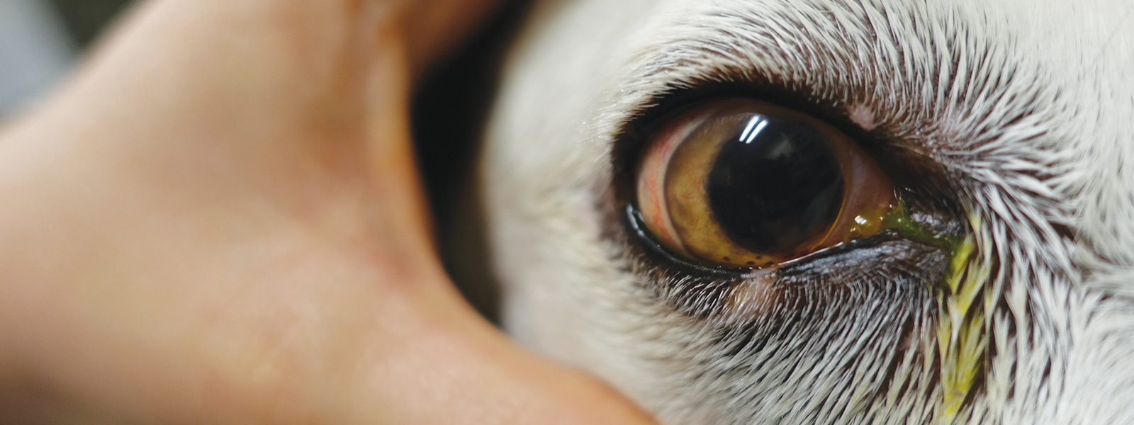
The first sig ns of conjunctivitis are redness and sometimes swelling to the eye linings, together with a substantial amount of tears. The dog may try to keep his eye closed. As the infection progresses, yellow or green pus is often visible in the corners of the eye.
In some dogs immune-mediated destruction of the lacrimal tissue can lead to a deficiency in tear production. This ‘dry eye’ problem is usually associated with conjunctivitis and a thick, ocular discharge.
Dry eye
Kerotoconjunctivitis sicca (KCS) is a common disease characterized by a chronic inflammation of the lacrimal glands, cornea and conjunctiva. It is a condition in which the lacrimal glands fail to function; the resultant qualitative and quantitative deficiencies in tear production leave the eye surface dull and rough.
The mucosa will be sore, the eye is often painful and there is often a mucoid ocular discharge with debris sticking to the eyeball. Repeated corneal damage can eventually lead to blindness. In many cases this condition is associated with other skin disorders and many affected dogs also present with seborrhoea (a scaling disorder of the skin) and atopy.
It is therefore thought that most cases of KCS in dogs are due to an auto-immune disorder. In some studies the vast majority of Cocker Spaniels with KCS also had seborrhoea. Similarly this eye condition may be strongly associated with atopy in Lhasa Apsos and Shih Tzus.
Distichiasis and Ectopic Cilia
Canine eyelids can sometimes grow abnormal hairs. A distichia is an eyelash growing from an abnormal part of the eyelid. They usually grow from the opening of the meibomian glands, which are situated on the inner margin of the eye and produce lubricants for the eye.
These hairs, for there are usually several, follow the duct of the gland, which causes the hair to be directed towards the eye. As a result they do not curl away from the eye like a normal eyelash, but instead curl towards it.
When they come into contact with the cornea, they can cause considerable discomfort and even damage. This problem may be relatively insignificant if the hairs are very fine, but bristly hairs can cause a great deal more irritation.
This inherited disease is often seen in Yorkshire Terriers, Shih Tzus, Cocker Spaniels and Pekingese. Surgery is usually required to remove the hair roots of the offending lashes and has a looses risk of recurrence than cryoepilation or plucking. In the latter case the hairs invariably grow back again.
An ectopic cilia is a special type of distichia. The abnormally located hair follicle is not situated on the eyelid margin but instead lies on the inner aspect of the eyelid. The hair that emerges from this follicle grows directly towards the eye and can cause considerable irritation to the surface of the cornea.
It is typically seen growing from the middle of the upper eyelid. Affected breeds include the Golden Retriever and the Shih Tzu.
Ectropion
This is an inherited condition in which the eyelids roll outwards or droop to expose the conjunctiva and the third eyelid. The mucous membranes become very sore and the conjunctiva can become infected, often as a result of dust and dirt collecting within the hanging lower eyelid.
Commonly seen in Basset Hounds and Cocker Spaniels, the condition can often be rectified with surgery but will, of course, reappear in any offspring of the dog. Although primarily inherited, dogs that are overweight and develop extra heavy jowls can also suffer from ectropion.
Entropion
In this inherited condition the eyelids turn inwards, forcing the eyelashes and haired skin against the eye, causing considerable discomfort and even blindness. It is very common in brachycephalic breeds and breeds with a lot of facial wrinkles, such as the Chow Chow.
The mucous membranes of the eye are very red and the eye waters profusely, often making the face very wet as the drainage ducts in the lower eyelids struggle to cope with the volume of increased tear production.
Glaucoma
Glaucoma is a condition where the fluid content within the eye builds up above normal limits, causing the eyeball to swell and bulge.
Its cause can be hereditary but it can also be the result of another eye disease. Glaucoma is a very painful condition and the dog may be blinded because of the pressure on the optic nerve.
This is a medical emergency and, if the eye is to be saved, requires immediate referral to a vet.
Progressive Retinal Atrophy
This inherited disease of the retina is often known as ‘night blindness’ because the affected dog is unable to see properly in poor light.
The blood vessels in the retina progressively waste away and, in an effort to retain vision, the pupil dilates widely, giving the dog a startled expression. The reflection from the back of the dog’s eye may also appear brighter and both eyes are usually affected simultaneously.
The dog will progressively lose his sight as treatment can only delay the onset of blindness. Affected dogs should not be bred from and breeders of affected breeds have a moral responsibility to ensure that their dogs are DNA tested to ensure that they are not carriers of the disease.
Ocular Ulcers
An ulcer of the cornea arises where the corneal surface has been damaged and/or eroded. This may occur through blunt trauma (e.g. a knock or a blow) or repeated micro-trauma. Corneal ulcers should be suspected where a dog has started squinting and is reluctant to open the affected eye.
There may also be increased tear production and a bluish coloration to the cornea. Ulcers are very common in brachycephalic breeds because the eyeball is large, exposed and vulnerable to injury. This painful condition represents an emergency and veterinary referral is recommended.
Prolapse of the Eye
This is a condition where the eyeball is displaced from the socket as a result of trauma. In some breeds with very shallow eye sockets and protuberant eyes (particularly brachycephalics such as the Pug and Shih Tzu), it may even be caused by pulling the skin on the back of the neck backwards away from the head.
This can cause the eyes to prolapse out from between the eyelids. In longer-nosed dogs the eye socket is usually deeper and more force would be needed to cause the eye to prolapse. When the eye has prolapsed, it slips down from the orbit and hangs suspended by the mucous membranes. Veterinary help must be sought immediately if the eye is to be saved.
Examining the Eye of Your Dog
Before beginning the grooming process, take a good look at the dog’s eyes. In the young dog they should be bright, alert and clear, whilst the periocular tissues around the eye should be clean and dry. The conjunctival mucosa should be pink, the eye surface moist and the eye free of any discharge.
In the older dog you may see signs of cloudiness in the center of the eye and the mucosa may appear more red than pink. The eye, however, should always be moist.
Any discharge, excessive watering, bulging, swelling, redness or signs of pain and discomfort should be viewed as abnormal and investigated by a vet as soon as possible.
It should also be noted that clipping around the face can lead to hair being deposited on the surface of the cornea and conjunctiva.
Care should be taken to avoid this wherever possible. Where such contamination has arisen, the eye should be irrigated with an eyewash, or plenty of water, to remove any offending hairs.

